Virus Replication

Summary

Many enveloped viruses are released from infected cells by maturing and budding at the plasma membrane. During this process, components of the viral core are incorporated into membrane vesicles that contain viral transmembrane proteins, called “spike” proteins. For many years, these spike proteins, which are required for infectivity, were believed to be incorporated into virions through direct interaction between their cytoplasmic domains and components of the viral core.

More recent evidence shows that while such direct interactions drive budding in alphaviruses, this may not be the case for negative-stranded RNA viruses and retroviruses. These viruses can generate particles in the absence of spike proteins, using only viral core components to drive the process. In some cases, the spike proteins, without the viral core, can be released as virus-like particles. Therefore, optimal sprouting and release may depend on a concerted ‘push and pull action of the core and spike, where oligomerization of both components plays a crucial role.

As viruses are obligate intracellular pathogens, they cannot replicate without the machinery and metabolism of a host cell. Although the replicative life cycle of viruses differs greatly between virus species and categories, there are six basic stages that are essential for viral replication.

1. Adhesion: Viral proteins on the capsid or phospholipid envelope interact with specific receptors on the host cell surface. This specificity determines the host range (tropism) of a virus.

2. Penetration: The process of binding to a specific receptor can induce conformational changes in the viral capsid proteins, or the lipid envelope, resulting in fusion of the viral and cellular membranes. Some DNA viruses can also enter the host cell through receptor-mediated endocytosis.

3. Stripping: The viral capsid is removed and degraded by viral enzymes or host enzymes that release the viral genomic nucleic acid.

4. Replication: After the viral genome has been unwrapped, transcription or translation of the viral genome begins. It is this stage of viral replication that differs greatly between DNA and RNA viruses and viruses with opposite nucleic acid polarity. This process culminates in the de novo synthesis of viral proteins and genome.

5. Assembly: After de novo synthesis of the viral genome and proteins, which can be modified post-transcriptionally, the viral proteins are packaged with the newly replicated viral genome into new virions that are ready to be released from the host cell. This process can also be called maturation.

6. Virion release: There are two methods of viral release: lysis or budding. Lysis results in the death of an infected host cell, these types of viruses are called cytolytic. One example is variola major, also known as smallpox. Enveloped viruses, such as the influenza A virus, are usually released from the host cell by budding. It is this process that results in the acquisition of the phospholipid viral envelope. These types of viruses do not usually kill the infected cell and are called cytopathic viruses.

After virion release, some viral proteins remain within the host cell membrane, serving as potential targets for circulating antibodies. Residual viral proteins that remain within the host cell cytoplasm can be processed and presented at the cell surface on MHC class I molecules, where they are recognized by T cells.

Materials And Methods

  • Viruses and cells.

Vero and HeLa cells were maintained in Dulbecco’s Modified Eagle’s Medium (Invitrogen) supplemented with 5% fetal bovine serum, 5% bovine serum, penicillin and streptomycin. Virus stocks were prepared by infecting confluent monolayers of Vero cells with the KOS strain of HSV type 1 or the Becker strain of pseudorabies virus (PRV) at a multiplicity of infection of 0.01 for 1 h.

Why envelope forms in some kind of the viruses only? What is enzyme and mechanism behind it to form an envelope? - Quora

After infection, cells were incubated in Dulbecco’s modified Eagle’s medium supplemented with 2% fetal bovine serum, 25 mM HEPES buffer, glutamine (0.3 μg/ml), penicillin, and streptomycin until a cytopathic effect was visualized. complete (~3 days). Cells were then scraped into the medium, frozen (-80 °C) and thawed (37 °C) three times, and then sonicated (4 °C) at moderate power for three pulses of 1 min. Cellular debris was removed by centrifugation and aliquots were frozen at -80°C until use.

  • Mutant viruses.

The GC-null mutant and revertant (GC-R) used in these studies were propagated in Vero cells as described previously (kindly provided by Harvey Friedman). The GB null mutants (KO82 and F-BAC GB-) and complementary cells (VB38) used for their growth were generously provided by David Johnson. To generate GB-deficient virions, the progeny of GB-depleted viruses were obtained after infection of non-complementary Vero cells. The ΔUL16 mutant used in the supporting studies (see Supplementary Material) was a gift from Joel Baines and has been described previously.

  • MEN treatment of herpesviruses.

Extracellular virions harvested between 18 and 24 h post-infection were treated with 10 mM NEM for 30 min at 37 °C, either before or after unwrapping with NP-40 (final concentration, 0.5 %. ; I3021; ​​Sigma). Previous work revealed that this concentration and treatment time efficiently modify free cysteines, and in our system also maintain the UL16-capsid interaction in virions.

After NEM treatment, virions and capsids were pelleted at 83,500 × g for 1 hour and samples were separated on 10% sodium dodecyl sulfate (SDS) polyacrylamide gels and electroblotted onto nitrocellulose membranes. The enhanced chemiluminescence method of immunoblot analysis was performed according to the manufacturer’s instructions (Amersham). Rabbit anti-HSV UL16 and anti-VP5 sera were used at dilutions of 1:6,000 and 1:7,500, respectively. PRV UL16-specific antibodies were kindly provided by Thomas Mettenleiter and used at a dilution of 1:15,000.

  • Virus-cell binding assay.

Confluent monolayers of cells in 100-mm dishes were incubated with 1 mL of virus stock (∼5 × 10⁸ PFU) at 4°C with rocking for 45 min to allow virus attachment. NEM was added to the virus before or after incubation with cells for 30 min at 4°C. Unbound virus and residual NEM were removed by washing the cells with 5 ml of phosphate-buffered saline (PBS).

Viral membranes that remained attached to the cells were solubilized with 4 mL of NP-40 lysis buffer (0.5% NP-40, 150 mM NaCl, 50 mM Tris-HCl [pH 8.0]) for 15 min. at 37°C.  Lysates were removed from the dish by pipetting, and insoluble material and cell debris were removed by centrifugation for 10 min at 3,829 × g. The supernatant capsids were pelleted through a 700 μl 30% sucrose cushion (wt/vol in TNE [20 mM Tris-HCl {pH 7.6}, 100 mM NaCl, 1 mM EDTA]) in a rotor. SW55ti at 83,500 × g for 1 h.

Pellets were dissolved in SDS sample buffer (2% SDS, 62.5 mM Tris-HCl, 10% glycerol, 0.025% bromophenol blue, 50 mM dithiothreitol [DTT], and 5% β-mercaptoethanol [BME]), boiled and separated by SDS. – polyacrylamide gel electrophoresis (PAGE) in 10% gels, and analyzed by immunoblotting using specific antibodies for VP5 and UL16. The amount of UL16 was determined by densitometry and normalized to VP5 levels.

Prion Diseases

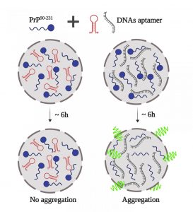
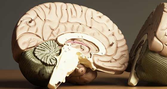
Prion, which is an abnormal form of a normally harmless protein found in the brain that is responsible for a variety of fatal neurodegenerative diseases in animals, including humans, is called transmissible spongiform encephalopathies.

In the early 1980s, American neurologist Stanley B. Prusiner and his colleagues identified the “infectious protein particle,” a name that was shortened to “prion” (pronounced “pree-on”). Prions can enter the brain through infection or can arise from mutations in the gene that codes for the protein.

Once present in the brain, prions multiply by inducing benign proteins to fold into an abnormal shape. This mechanism is not fully understood, but another protein normally found in the body may also be involved. Normal protein structure is thought to consist of a series of flexible coils called alpha-helices. In prion protein, some of these helices are stretched out into flat structures called beta-strands.

The normal conformation of the protein can be degraded quite easily by cellular enzymes called proteases, but the prion protein form is more resistant to this enzymatic activity. Therefore, as prion proteins multiply, they are not broken down by proteases and instead accumulate inside neurons, destroying them. The progressive destruction of neurons eventually causes brain tissue to fill with holes in a sponge-like or spongiform pattern.

Prion diseases that affect humans include: Creutzfeldt-Jakob disease, Gerstmann-Sträussler-Scheinker disease, fatal familial insomnia, and kuru. Prion diseases that affect animals include scrapie, bovine spongiform encephalopathy (commonly called mad cow disease), and chronic wasting of mule deer and elk.

For decades, doctors thought these diseases were the result of infection with slow-acting viruses, named for the long incubation times required for the diseases to develop. These diseases were called, and are sometimes still called slow infections. The pathogen of these diseases has certain viral attributes, such as extremely small size and strain variation, but other properties are atypical of viruses. In particular, the agent is resistant to ultraviolet radiation, which normally inactivates viruses by destroying their nucleic acid.

Prions differ from all other known disease-causing agents in that they appear to lack nucleic acid, that is, DNA or RNA, which is the genetic material that all other organisms contain. Another unusual feature of prions is that they can cause hereditary, infectious and sporadic diseases; for example, Creutzfeldt-Jakob disease manifests itself in all three forms, with sporadic cases being the most common.

Prion proteins can act as infectious agents, spreading disease when transmitted to another organism, or they can arise from an inherited mutation. Prion diseases also show a sporadic pattern of incidence, meaning that they appear to appear randomly in the population. The underlying molecular process that causes the prion protein to form in these cases is unknown. Other neurodegenerative disorders, such as Alzheimer’s disease or Parkinson’s disease, may arise from molecular mechanisms that result in protein misfolding that are similar to those that cause prion diseases.

Types of prion diseases

Prion disease can occur in both humans and animals. Below are some different types of prion diseases.

Human prion diseases

  • Creutzfeldt-Jakob disease (CJD). First described in 1920, CJD can be acquired, inherited, or sporadic. Most CJD cases are sporadic.
  • Variant Creutzfeldt-Jakob disease (vCJD). This form of CJD can be acquired by eating the contaminated meat of a cow.
  • Fatal familial insomnia (FFI). FFI affects the thalamus, which is the part of your brain that manages your sleep-wake cycles. One of the main symptoms of this condition is worsening insomnia. The mutation is dominantly inherited, meaning an affected person has a 50 per cent chance of passing it on to their children.
  • Gerstmann-Straussler-Scheinker syndrome (GSS). GSS is also inherited. Like FFI, it is dominantly transmitted. It affects the cerebellum, which is the part of the brain that handles balance, coordination, and equilibrium.
  • Kuru: Kuru was identified in a group of people from New Guinea. The disease was transmitted through a form of ritual cannibalism in which the remains of deceased relatives were consumed.

Risk factors for these diseases include:

  • Genetics. If someone in your family has an inherited prion disease, you are also at higher risk of having the mutation.
  • Years. Sporadic prion diseases tend to develop in older adults.
  • Animal products. Eating animal products that are contaminated with a prion can give you a prion disease.
  • Medical procedures. Prion diseases can be transmitted through contaminated medical equipment and nerve tissue. Cases, where this has happened, include transmission through contaminated corneal transplants or dura mater grafts.

What are the symptoms of prion disease?

Prion diseases have very long incubation periods, often on the order of many years. When symptoms develop, they get progressively worse, sometimes rapidly. Common symptoms of prion disease include:

  • difficulties with thinking, memory, and judgment
  • personality changes such as apathy, agitation, and depression
  • confusion or disorientation
  • involuntary muscle spasms (myoclonus)
  • loss of coordination (ataxia)
  • difficulty sleeping (insomnia)
  • difficult or slurred speech
  • vision problems or blindness

How is prion disease treated?

There is currently no cure for prion disease. But treatment focuses on providing supportive care. Examples of this type of care include:

  • Medications: Some medications may be prescribed to help treat symptoms. Examples include:

– reduce psychological symptoms with antidepressants or sedatives
– providing pain relief using opiate medications
– relieve muscle spasms with drugs such as sodium valproate and clonazepam

  • Assistance. As the disease progresses, many people need help to care for themselves and do their daily activities.
  • Providing hydration and nutrients. In the advanced stages of the disease, intravenous fluids or a feeding tube may be required.

Malaria

Overview

Malaria is a disease caused by a parasite. The parasite is transmitted to humans through the bites of infected mosquitoes. People who have malaria usually feel very sick with a high fever and chills. Although the disease is rare in temperate climates, malaria remains common in tropical and subtropical countries. Every year, almost 290 million people become infected with malaria and more than 400,000 people die from the disease.

To reduce malaria infections, global health programs distribute preventive medicines and insecticide-treated nets to protect people from mosquito bites. The World Health Organization has recommended a malaria vaccine for use in children living in countries with a high number of malaria cases.

Protective clothing, mosquito nets, and insecticides can protect you while you travel. You can also take preventive medicine before, during, and after a trip to a high-risk area. Many malaria parasites have developed resistance to the common drugs used to treat the disease.

Symptoms

Malaria signs and symptoms may include:

  • Fever
  • Shaking chills
  • General feeling of discomfort.
  • Headache
  • nausea and vomiting
  • Diarrhea
  • Abdominal pain
  • muscle or joint pain
  • Fatigue
  • Fast breathing
  • fast heart rate
  • Cough

Some people who have malaria experience cycles of malaria “attacks.” An attack usually begins with chills and chills, followed by a high fever, sweats, and a return to normal temperature. The signs and symptoms of malaria usually begin a few weeks after being bitten by an infected mosquito. However, some types of malaria parasites can remain dormant in your body for up to a year.

When to see a doctor

Talk to your doctor if you experience a fever while living in or after travelling to a high-risk malaria region. If you have severe symptoms, seek emergency medical attention.

Causes

Malaria is caused by a unicellular parasite of the genus Plasmodium. The parasite is most commonly transmitted to humans through mosquito bites.

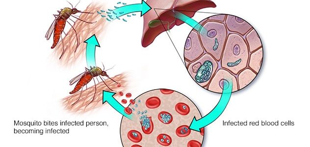
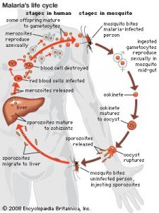
Mosquito transmission cycle

  • Uninfected mosquito. A mosquito becomes infected by feeding on a person who has malaria.
  • Transmission of the parasite. If this mosquito bites you in the future, it can transmit malaria parasites to you.
  • In the liver. Once the parasites enter your body, they travel to your liver, where some types can lie dormant for up to a year.
  • In the bloodstream. When the parasites mature, they leave the liver and infect red blood cells. This is when people usually develop malaria symptoms.
  • To the next person. If you are bitten by an uninfected mosquito at this point in the cycle, it will become infected with malaria parasites and can pass them on to other people it bites.

Other modes of transmission

Because the parasites that cause malaria affect red blood cells, people can also get malaria from exposure to infected blood, including:

  • From mother to unborn child
  • Through blood transfusions.
  • By sharing needles used to inject drugs

Risk factor’s

The biggest risk factor for developing malaria is living in or visiting areas where the disease is common. These include the tropical and subtropical regions of:

  • Sub-Saharan Africa
  • South and Southeast Asia
  • pacific islands
  • Central America and northern South America

The degree of risk depends on local malaria control, seasonal changes in malaria rates, and the precautions you take to avoid mosquito bites.

Risks of a more serious illness

People at higher risk for serious illness include:

  • toddlers and babies
  • Older adults
  • Travelers from malaria-free areas
  • Pregnant women and their unborn children

In many countries with high rates of malaria, the problem is compounded by a lack of access to preventive measures, medical care and information.

Immunity may decrease

Residents of a region with malaria may be exposed enough to the disease to acquire partial immunity, which can lessen the severity of malaria symptoms. However, this partial immunity can disappear if you move to a place where you are no longer frequently exposed to the parasite.

Complications

Malaria can be fatal, particularly when caused by Plasmodium species common in Africa. The World Health Organization estimates that around 94% of all malaria deaths occur in Africa, most commonly in children under 5 years of age. Malaria deaths are usually related to one or more serious complications, including:

  • Cerebral malaria. If parasite-filled blood cells block small blood vessels in the brain (cerebral malaria), brain swelling or brain damage can occur. Cerebral malaria can cause seizures and coma.
  • Respiratory problems. Fluid built up in the lungs (pulmonary oedema) can make it hard to breathe.
  • Organ failure. Malaria can damage the kidneys or liver or cause the spleen to rupture. Any of these conditions can be life-threatening.
  • Anaemia. Malaria can cause you do not to have enough red blood cells to adequately supply your body’s tissues with oxygen (anaemia).
  • Low blood sugar. Severe forms of malaria can cause low blood sugar (hypoglycemia), as can quinine, a common medicine used to combat malaria. Very low blood sugar can cause coma or death.

Malaria may reappear

Some varieties of the malaria parasite, which usually cause milder forms of the disease, can persist for years and cause relapses.

Prevention

If you live in or travel to an area where malaria is common, take steps to avoid mosquito bites. Mosquitoes are most active between dusk and dawn. To protect yourself from mosquito bites, you should:

  • Cover your skin. Wear pants and long-sleeved shirts. Put on your shirt and tuck your pant legs into your socks.
  • Apply insect repellent to the skin. Use an insect repellent registered with the Environmental Protection Agency on any exposed skin. These include repellents that contain DEET, picaridin, IR3535, oil of lemon eucalyptus (OLE), para-menthane-3,8-diol (PMD), or 2-undecanoate. Do not use a spray directly on your face. Do not use products with OLE or PMD in children under 3 years of age.
  • Apply repellent to clothing. Sprays containing permethrin are safe to apply to clothing.
  • Sleep under a net. Bed nets, particularly those treated with insecticides such as permethrin, help prevent mosquito bites while you sleep.

Preventive medicine

If you’re travelling to a place where malaria is common, talk to your doctor a few months beforehand about whether you should take medication before, during, and after your trip to help protect you from malaria parasites. In general, the medicines taken to prevent malaria are the same ones used to treat the disease. The medicine you take depends on where and how long you travel and on your own health.

Vaccine

The World Health Organization has recommended a malaria vaccine for use in children living in countries with a high number of malaria cases. Researchers continue to develop and study malaria vaccines to prevent infection.